Mammograms and Breast Health
What is a mammogram?
A mammogram is an X-ray image of your breast. It's used to find and diagnose breast disease. A mammogram may be done if you have breast problems such as a lump, pain, or nipple discharge. A mammogram is also done as a screening test if you don’t have breast problems. It can check for breast cancers, noncancerous or benign tumors, and cysts before they can be felt.
A mammogram can’t prove that an abnormal area is cancer. If a mammogram shows an area in your breast that is abnormal or may be cancer, follow up tests such as a breast ultrasound exam or an MRI may be done. For a conclusive diagnosis your healthcare provider removes a sample of breast tissue (biopsy) by needle or during surgery. The tissue will be examined at a lab under a microscope to see if it's cancer.
A mammogram uses a low dose of radiation.
What are the different types of mammograms?
There are 2 types of mammograms:
-
Screening mammogram. This is used to find any breast changes in people who have no signs of breast cancer. Often 2 X-rays are taken of each breast. A mammogram can find a tumor before it can be felt.
-
Diagnostic mammogram. This is used to diagnose abnormal breast changes. These may include a lump, pain, nipple thickening or discharge, or a change in breast size or shape. More pictures are taken than during a screening mammogram. A diagnostic mammogram is also used to check any problems found on a screening mammogram.
How is a mammogram done?
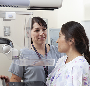
X-rays of the breast are different from X-rays for other parts of your body. The breast is squeezed, or compressed, by the mammogram equipment. This spreads the breast tissue apart. Because of this, the radiation dose is lower and there is a better picture of your breast tissue. You may feel some mild pain when your breast is compressed. The compression only lasts for a few seconds for each image of your breast. An X-ray technologist takes the X-rays. The films are read by a radiologist. The results are given to your healthcare provider.
Mammograms are currently done with the help of a computer to make digital images. Digital mammograms are done the same way as a standard mammogram.

What conditions does a mammogram show?
A mammogram can show the following conditions:
Calcifications are tiny mineral deposits in the breast tissue. There are 2 types of calcifications:
-
Macrocalcifications. These are larger calcium deposits that often mean worsening changes in the breast. These changes may include aging of the breast arteries, past injuries, swelling, or inflammation.
-
Microcalcifications. These are tiny (less than 1/50 of an inch) specks of calcium. When many microcalcifications are seen in one area, they are called a cluster.
Masses may happen with or without calcifications. Masses may be caused by:
-
A cyst. This is a noncancer (benign) collection of fluid in the breast. It can’t be diagnosed by a physical exam alone or by a mammogram alone. A breast ultrasound or aspiration with a needle is needed. If a mass is not a cyst, you may need more tests.
-
Benign breast conditions. Some masses can be checked with regular mammograms. But for others you may need a biopsy.
-
Breast cancer
Who should get a screening mammogram?
Different health experts have different recommendations for people who have no symptoms of breast cancer:
-
The U.S. Preventive Services Task Force recommends screening mammograms every 2 years for women age 40 through 74. This applies to people at average risk of breast cancer.
-
The American Cancer Society recommends screening be a choice for those at average risk, starting at age 40. Mammograms should be done every year for everyone ages 45 to 54. Then you can switch to mammograms every 2 years. Or you have the choice to continue annual mammograms. Screening can continue as long as a woman is in good health and expected to live 10 years or more.
Talk with your healthcare provider to find out which screening guidelines are right for you.
If you are at higher risk for breast cancer, talk with your provider about:
-
Starting screening mammograms earlier
-
Having additional tests such as a breast ultrasound or MRI
-
Having mammograms more often